Feburary, 2017
In Japan, “PALFIQUE™ UNIVERSAL FLOW” is sold under the name “ESTELITE UNIVERSAL FLOW™”, and “PALFIQUE™ Bond” is sold under the name “Tokuyama Bond Force™ II”.
Composite resin comes in two consistencies: paste and flowable. In my daily clinical practice, I have mostly used the paste type, which offers superior mechanical properties and sculptability along with a wide selection of shades. Conventional flowable resin, by contrast, often fails to match the tooth shade on its own, tends to string during placement, and offers limited viscosity options. Consequently, I used it only in limited situations, such as Class V cavities and wedge-shaped defects, lining markedly uneven cavity floors, filling small cavities and fine details, and filling areas difficult to access with instruments, all with the aim of preventing air bubbles and shortening chair time. However, the newly released ESTELITE UNIVERSAL FLOW™ offers three different viscosities (Super Low, Medium, and High), making it suitable for a wide range of cavity forms and allowing practitioners to choose their preferred viscosity. The material also does not string during placement, and its high light diffusion improves shade matching, allowing stress-free use in terms of both handling and esthetics. This product has also enabled me to use flowable resin in Class IV and Class II cavities, where I previously found it difficult to use.
This article, therefore, introduces the use of ESTELITE UNIVERSAL FLOW™ in clinical cases involving the anterior teeth and premolars.
Case 1: Filling a Class II Premolar Cavity (High and Medium)
Before treatment, #44 had a metal inlay. Removing the inlay and caries revealed a mesiodistally narrow proximal cavity, where a paste resin would have been difficult to pack with sufficient pressure and would most likely have left the margins underfilled, so a flowable resin, which can be dispensed through a fine nozzle, was used. With postoperative esthetics in mind, the A2 shade of High was used on the proximal surface to avoid a darkened result, while the shallow occlusal cavity was filled with the CE shade of Medium.
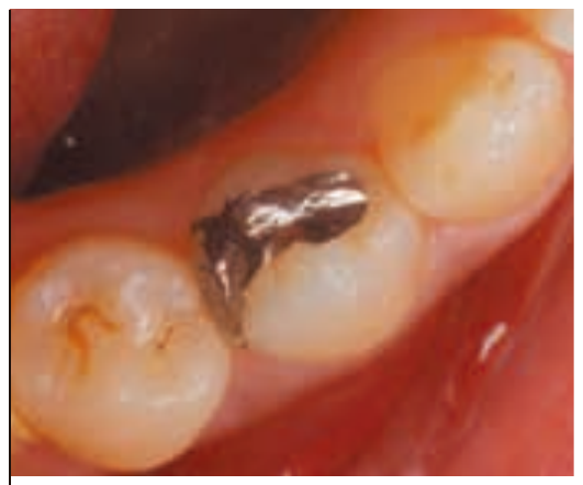
Fig. 1 Preoperative condition.
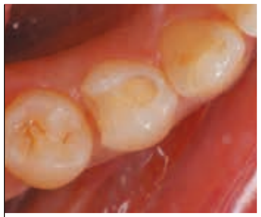
Fig. 2 The metal inlay and caries were removed.
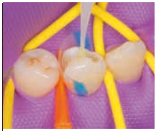
Fig. 3 Bonding was performed using Tokuyama Bond Force™ II.
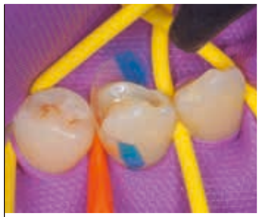
Fig. 4 The surface was dried with medium-pressure air for about five seconds.
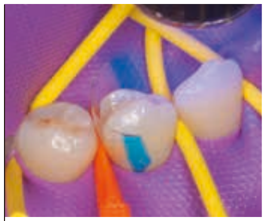
Fig. 5 Light curing was performed for 10 seconds.
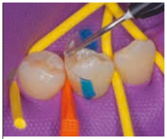
Fig. 6 The proximal surface was filled using ESTELITE UNIVERSAL FLOW™ High (A2).
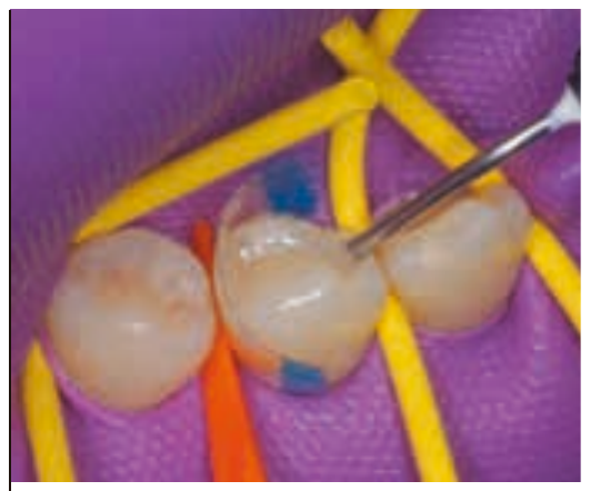
Fig. 7 Since the occlusal cavity was shallow, it was filled with ESTELITE UNIVERSAL FLOW™ Medium (CE).
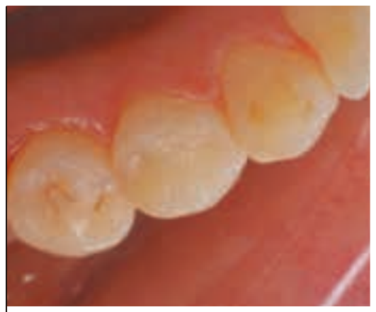
Fig. 8 After contouring and polishing, excellent shade matching could be seen.
Case 2: Filling the Cervical Region of Anterior Teeth (High, Medium, and Super Low)
The patient presented with a chief complaint of esthetic dissatisfaction with anterior composite resin fillings placed three years earlier. For tooth #11, where the cavity was confined to enamel above the gingival margin, the CE shade of ESTELITE UNIVERSAL FLOW™ Medium was used. For the cervical area, from the gingival crest to below the gingival margin, the high-chroma A4 shade of ESTELITE UNIVERSAL FLOW™ Super Low was used while the A3 shade was used for the proximal surfaces. Layering different shades of flowable resin enables the creation of natural-looking restorations.
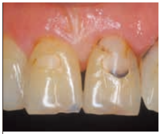
Fig. 1 Preoperative condition. A brown line is visible along the composite resin on #11, and caries are present on #21 and #22.
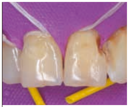
Fig. 2 The existing composite resin and caries were removed.
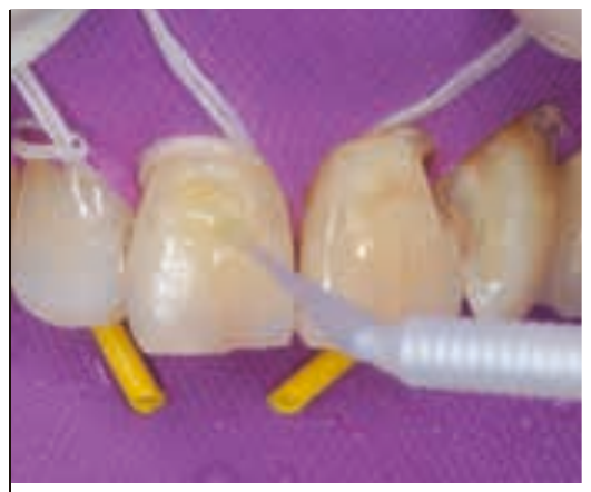
Fig. 3 Tokuyama Bond Force II was applied, dried and light-cured.
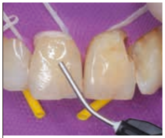
Fig. 4 Since the cavity on #11 was shallow (within enamel) and free of staining, it was filled with ESTELITE UNIVERSAL FLOW™ Medium (CE).
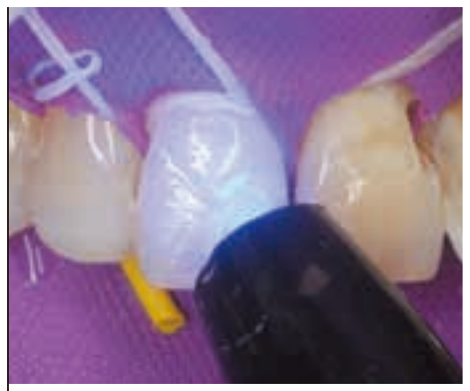
Fig. 5 Light curing was performed for 10 seconds.
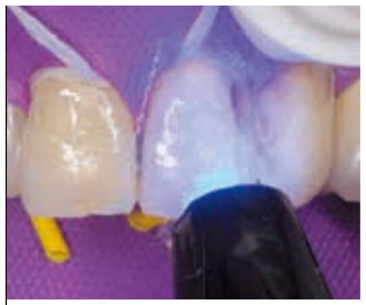
Fig. 6 ESTELITE UNIVERSAL FLOW™ High (A3) was used on the proximal surfaces between #21 and #22, and ESTELITE UNIVERSAL FLOW™ Super Low (A4) was used at the cervical area.
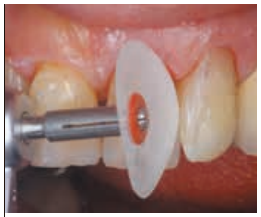
Fig. 7 Contouring was performed with a disc, paying careful attention to the form of the labial line angles.
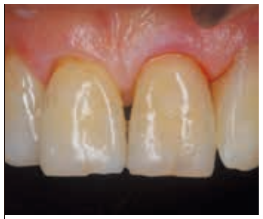
Fig. 8 Immediately postoperative.