May, 2017
In Japan, “PALFIQUE™ UNIVERSAL FLOW” is sold under the name “ESTELITE UNIVERSAL FLOW™”, and “PALFIQUE™ Bond” is sold under the name “Tokuyama Bond Force™ II”.
Because composite resin inherently shrinks during polymerization, contraction gaps can form. Consequently, composite fillings are regarded as technique-sensitive and prone to postoperative pain and secondary caries. For this reason, metal inlay restorations remain a common treatment for molar caries today.
In recent years, however, the physical properties of flowable composite resins themselves and the bond strength of bonding agents to tooth structure have improved remarkably. Therefore, I believe that adhesive restorative treatments, when performed correctly, can outperform indirect metal restorations, though the indications must be carefully assessed.
The new ESTELITE UNIVERSAL FLOW™ from Tokuyama Dental comes in three different viscosities. I would like to present my own clinical cases demonstrating a layered filling technique that utilizes each viscosity where it performs best within a single cavity. By combining layers with different levels of light transmission and value, this approach overcomes the functional weakness of composite resins while delivering highly esthetic results.
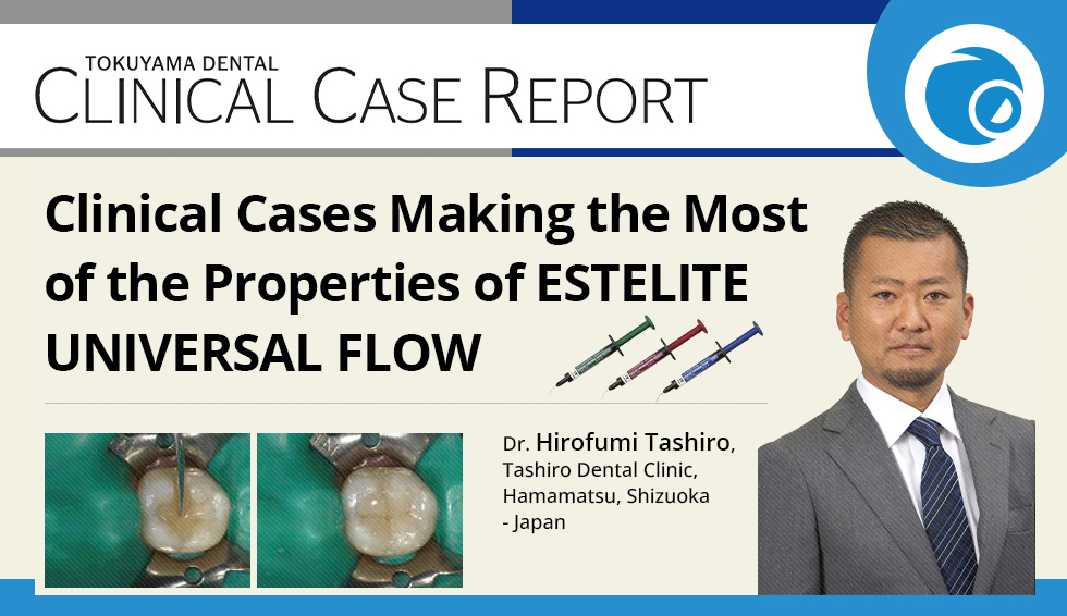
Case 1: Filling a Class I Molar Cavity (High, Medium, and Super Low)
Secondary caries was found on a mandibular first molar that had previously been filled with composite resin, and retreatment was required.
Here, I present a step-by-step technique aimed at achieving both esthetics and function. The OPA2 shade of High was used for the lining, the A3.5 shade of Medium for the dentin layer, and the A2 shade of Super Low for the enamel layer; this incremental approach was utilized to avoid the contraction gaps characteristic of composite resin fillings.
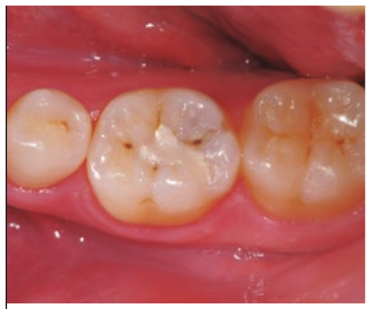
Fig. 1 Preoperative condition
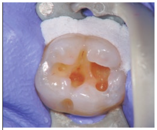
Fig. 2 After rubber dam isolation, the old filling and infected dentin are removed.
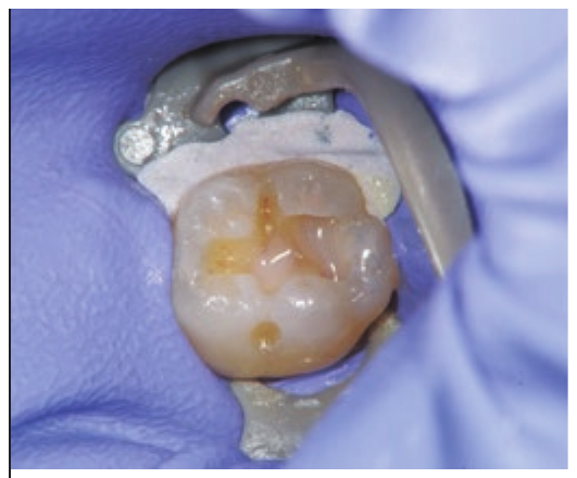
Fig. 3 Lining with High (OPA2) after bonding with Tokuyama Bond Force™ II.
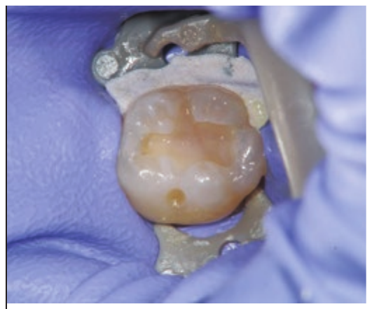
Fig. 4 The dentin layer is filled with Medium (A3.5).
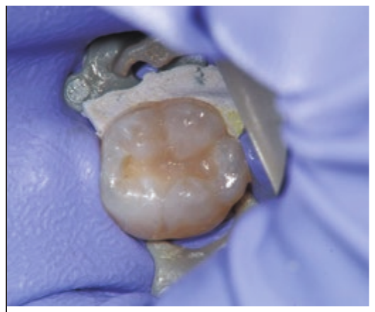
Fig. 5 The cusps are built up with Super Low (A2), working from the fossa toward the ridges.
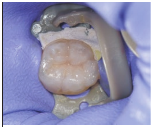
Fig. 6 Occlusal anatomy formed with Super Low (A2).
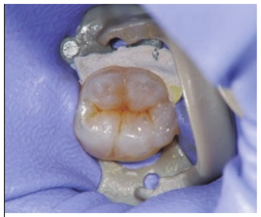
Fig. 7 The pits and fissures are stained using ESTELITE™ COLOR (DBr).
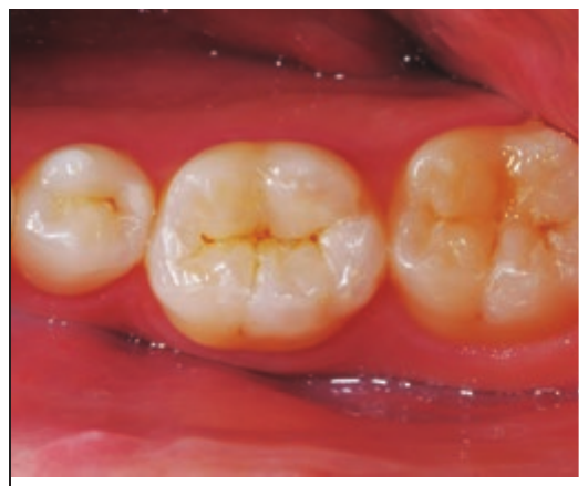
Fig. 8 Postoperative condition (10 days) following occlusal adjustment and polishing.
Case 2: Filling an Anterior Proximal Cavity (High, Medium, and Super Low)
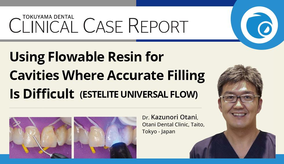
Treatment was planned after caries was found on the distal surface of a maxillary central incisor and a poorly fitting composite resin filling on the mesial surface of the lateral incisor.
Since flossing is essential for the patient’s self-care and maintenance after treatment, the composite resin filling must fit with a high degree of precision.
In Case 2, I present a step-by-step technique using a pre-curved matrix secured with a wedge. Flowable resins of different viscosities and light transmission are combined to achieve both close adaptation to the cavity walls and an esthetic result.
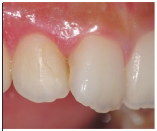
Fig. 1 Preoperative condition
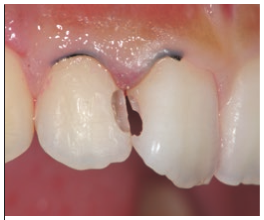
Fig. 2 The old filling and infected tooth structure are removed while preserving as much sound structure as possible.
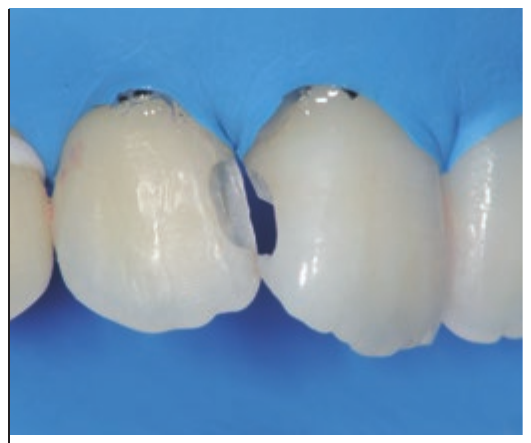
Fig. 3 Rubber dam isolation is performed after cavity preparation.
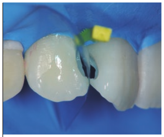
Fig. 4 After bonding with Tokuyama Bond Force™ II, the matrix is inserted at the proximal surface and secured with a wedge.
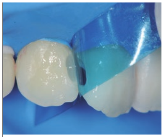
Fig. 5 Lining with High (OPA2) to improve adaptation to the small space formed between the matrix and the tooth structure.
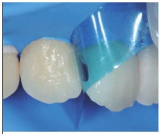
Fig. 6 Medium (OPA2), which has a lower viscosity, is used for the middle layer to keep the shade from washing out due to excessive light transmission through the proximal area.
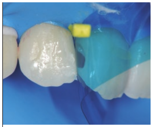
Fig. 7 For the outermost layer, Super Low (A2), which does not slump, is used to build up volume at the mesial marginal ridge. The distal surface of the central incisor is filled following the same steps.
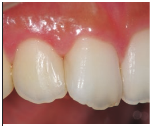
Fig. 8 Postoperative condition (7 days) following contouring and polishing.